In metabolic research, one finding has puzzled scientists for years. Drugs that activate the glucose-dependent insulinotropic polypeptide receptor, known as the GIP receptor, produce meaningful weight loss when combined with a separate appetite-regulating peptide. So do drugs that block the same receptor. Both effects show up in animal studies and in human clinical trials. That is a strange result. Normally, if flipping a switch one way causes an effect, flipping it the other way should reverse that effect, not replicate it.
A study published in Nature Metabolism now offers a detailed explanation. Researchers used genetically engineered mice to delete GIP receptors in specific brain areas and then tested how those mice responded to GIP-related compounds alone and in combination with other peptides. What they found is that the GIP receptor is doing two different jobs in two different parts of the brain, and each job is handled by a distinct anatomical location.
The GIP receptor paradox
The GIP receptor sits at the center of one of the more productive areas in recent metabolic research. Molecules that activate both the GIP receptor and the GLP-1 receptor simultaneously have shown strong effects on body weight and blood sugar in clinical settings. Researchers noticed, though, that the picture was more complicated than expected.
When a GIP receptor agonist, meaning a compound that turns the receptor on, is added to a GLP-1 receptor agonist, subjects lose more weight than they do on the GLP-1 receptor agonist alone. When a GIP receptor antagonist, meaning a compound that blocks the receptor, is added to the same GLP-1 receptor agonist, the result is also additional weight loss. Both combinations produce the same directional outcome even though the first turns the GIP receptor up and the second turns it off.
The authors of the Nature Metabolism paper describe this as an incompletely understood phenomenon and set out to map which brain structures account for each arm of the paradox.
Removing receptors from one region at a time
The research team used a technique called conditional knockout, which allows scientists to delete a specific gene in a targeted tissue while leaving the rest of the body untouched. They focused on two brain regions: the area postrema, a small structure near the base of the brainstem that sits outside the blood-brain barrier and is known to sense circulating signals in the bloodstream, and the hypothalamus, an older and larger structure involved in long-term energy balance and hunger.
One group of mice had GIP receptors removed only from the area postrema. A second group had GIP receptors removed only from the hypothalamus. Both groups were then exposed to GIP receptor agonists, GIP receptor antagonists, and a GLP-1 receptor agonist called liraglutide, individually and in combination. The researchers measured body weight, food intake, and behavioral responses to each treatment.
The area postrema handles direct appetite suppression
Mice without GIP receptors in the area postrema showed a reduced response when given acyl-GIP, an active form of the GIP peptide that functions as a GIP receptor agonist. Specifically, those mice ate more after treatment than control mice did, suggesting the area postrema is responsible for the immediate appetite-suppressing signal that GIP receptor agonism produces.
There was another revealing finding in this group. Peptide YY is a gut hormone associated with feelings of fullness, but at certain doses it also triggers an avoidance response in rodents, meaning the animals learn to avoid a flavor or location they associate with the uncomfortable sensation. Acyl-GIP normally reduces that avoidance behavior in control mice, a sign it may counteract some of the aversive signaling from peptide YY. In area postrema knockout mice, acyl-GIP lost that ability.
These mice also showed partial protection against diet-induced obesity compared with standard controls, which the authors interpret as a sign the area postrema GIP receptor is genuinely involved in the body weight response under high-fat dietary conditions. Importantly, when liraglutide was given to these mice, the weight loss response was similar to that seen in normal mice, confirming that the area postrema GIP receptor is specific to GIP-driven appetite effects and does not broadly alter how GLP-1 receptor agonism works.
The hypothalamus governs the synergy with other peptides
The hypothalamus knockout mice told a different story. Removing GIP receptors from the hypothalamus did not impair the appetite-suppressing effect of acyl-GIP, confirming that job belongs to the area postrema. What the hypothalamus knockout did affect was the interaction between GIP receptor antagonism and liraglutide.
In normal mice, adding a GIP receptor antagonist to liraglutide produces extra weight loss beyond what liraglutide achieves on its own. In hypothalamus knockout mice, that synergistic effect disappeared entirely. The researchers describe this as abolishing the additional weight loss conferred by GIP receptor antagonism when it is combined with GLP-1 receptor agonism.
The team also looked at whether nucleus tractus solitarius preproglucagon neurons, a cell population in the brainstem that is implicated in GLP-1 receptor biology, might be the link. They were not. The synergistic effect of hypothalamic GIP receptor removal appeared to operate through a separate pathway.
The hypothalamus knockout mice also showed an enhanced weight loss response to liraglutide alone compared with control mice, which the authors note is consistent with the idea that GIP receptor signaling in the hypothalamus normally acts as a kind of counterweight to GLP-1-driven weight loss.
Connection to amylin-related signaling
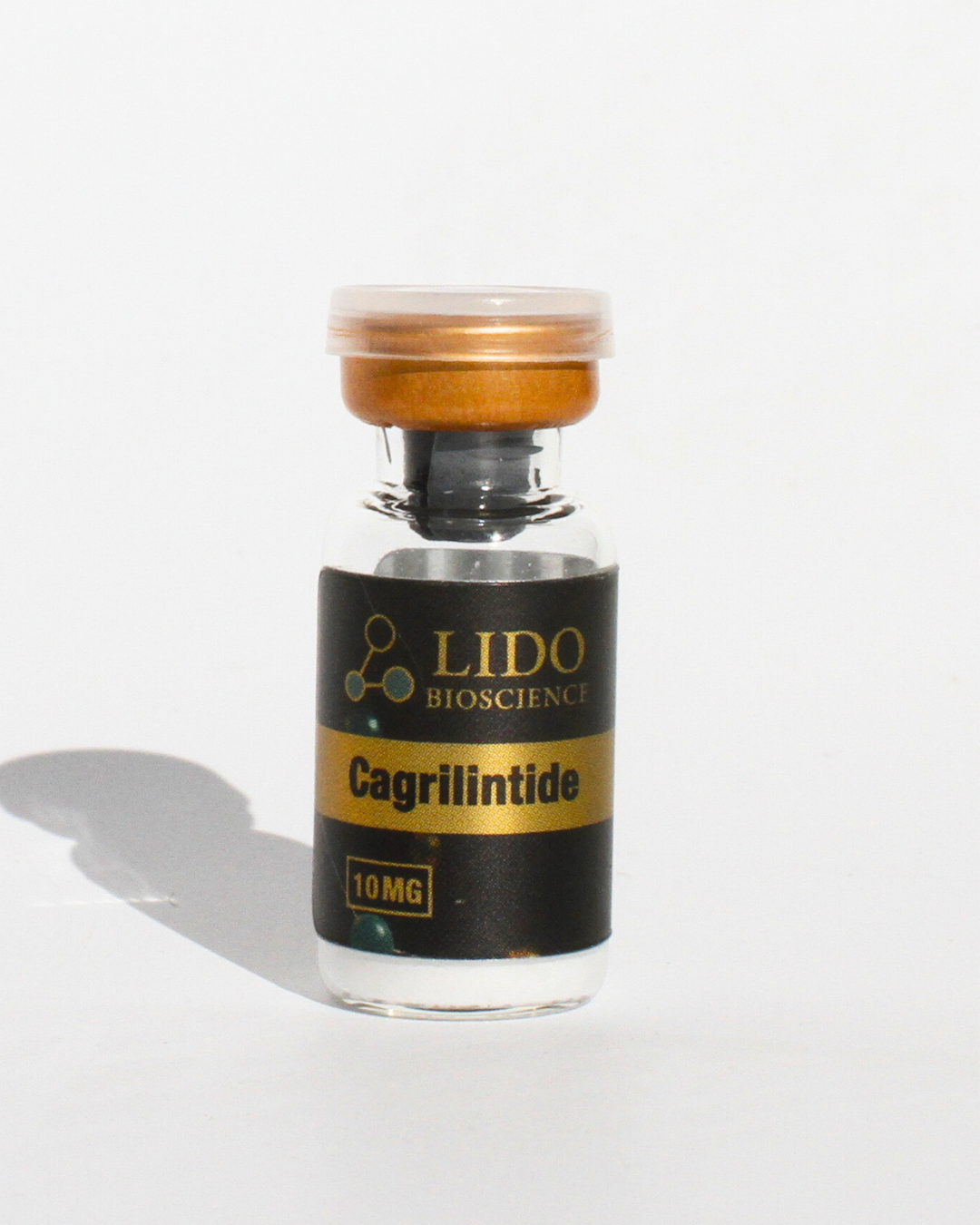
The study also examined how these brain-specific GIP receptor deletions interact with cagrilintide, a long-acting analogue of amylin, a peptide that is co-secreted with insulin and plays a role in satiety signaling. Both hypothalamus knockout mice and mice treated with a GIP receptor antagonist showed increased sensitivity to cagrilintide-induced weight loss compared with controls.
This finding extends the paradox in an interesting direction. It suggests that whatever the hypothalamic GIP receptor is doing when it is blocked or genetically removed, it creates conditions that make the body more responsive to amylin-pathway signaling. The mechanistic details of that interaction are not yet fully described, but the authors treat this as evidence that the hypothalamus is a hub where multiple metabolic peptide signals converge and interact.
What the findings mean for research
The study draws a reasonably clear map. The area postrema uses GIP receptor activation to suppress appetite acutely, and that is why GIP receptor agonists reduce food intake. The hypothalamus uses GIP receptor signaling in a way that modulates how the body responds to GLP-1 receptor and amylin receptor agonists, and blocking that signal actually amplifies the weight loss those compounds produce.
That map explains why both turning the GIP receptor on and turning it off can lead to weight loss. They are triggering different circuits in different locations. The agonist engages the brainstem detector. The antagonist releases a brake in the hypothalamus that would otherwise limit how much GLP-1 and amylin signals can drive weight loss.
The research was conducted in mice, and translating findings from rodent brain circuits to human physiology always requires caution. Still, the study provides a framework for understanding why combinations involving GIP receptor modulation continue to produce results in clinical research, and it identifies specific neural substrates that future studies can target to probe these mechanisms further.